Developmental Neurotoxicity (DNT) Testing

We are using multicellular and multiplexed human-based 2D/3D in vitro systems to evaluate chemicals regarding their potential to disturb brain development and cause developmental neurotoxicity.

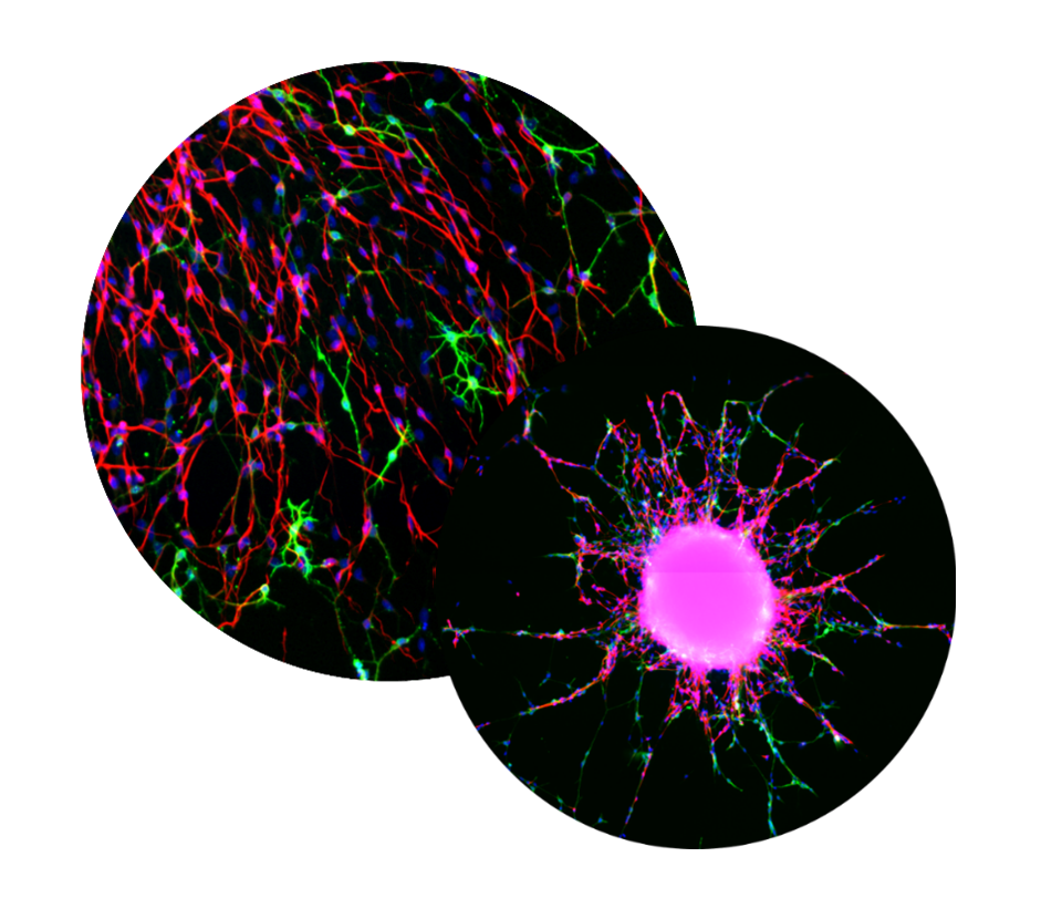
The so-called ‘Neurosphere Assay’ is comprised of a set of multicellular test methods that are based on primary human neural progenitor cells (hNPC). These cells are cultivated as three-dimensional floating or plated adhering spheres that can be used to study several key neurodevelopmental processes:

NPC proliferation is a fundamental neurodevelopmental process that, when disturbed, like in Zika virus-infected primary NPC, leads to microcephaly in children. The proliferation of primary hNPCs of fetal origin or human induced pluripotent stem cells (hiPSCs) grown as neurospheres in 3D is studied by measuring an increase in sphere size over time and/or by measuring DNA synthesis as BrdU incorporation in vitro.
Developmental Neurotoxicity (DNT) Testing
migration Assays

Cortex development takes place during the fetal phase of development. It involves radial glia migration leading to the development of a scaffold that is subsequently used by neurons to migrate and reach their final cortical destination. Thus, NPC migration is a fundamental neurodevelopmental process that, when disturbed, e.g. in methylmercury exposed children, leads to alterations in cortex development.

3D primary hNPCs of fetal origin are plated onto laminin-coated surfaces. Single NPCs radially migrate out of the sphere, thereby displaying radial glia morphology. It can be assessed by brightfield microscopy in unstained cells because radial glia build the scaffold for the other cell types’ migration and form the leading migration edge.

Control

DNT positive compound

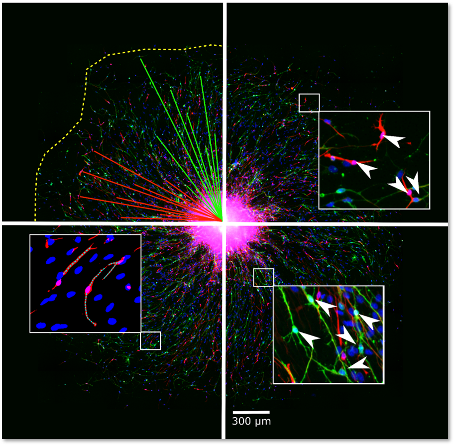
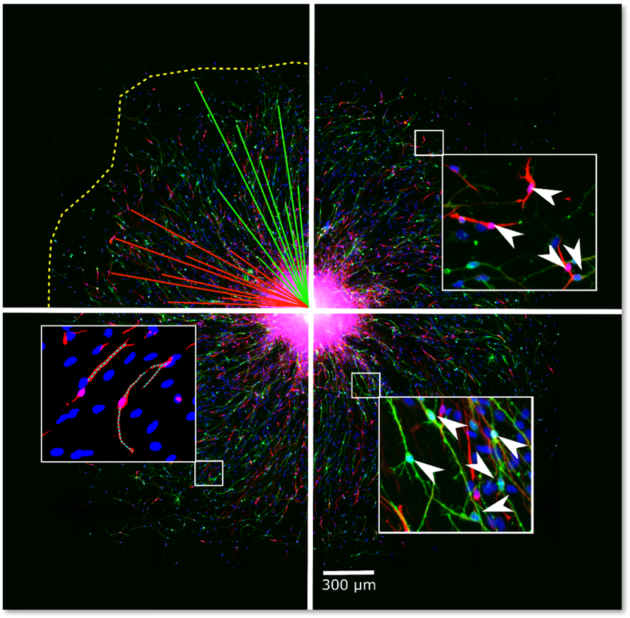
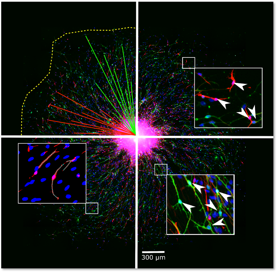
Secondarily, after radial glia differentiation, neurons and oligodendrocytes arise. In this multicellular secondary 3D model with the help of a convolutional neural network (CNN) immunostained neurons (TUBB3) and oligodendrocytes (O4) are identified and their position in the NPC migration area detected using our in-house developed software ‘Omnisphero’.

Migration Assays

The production of neurons is one of the prerequisites for proper brain development. During NPC migration out of the plated neurosphere cells differentiate into neurons, astrocytes and oligodendrocytes over a period of one to five days in vitro. Neuronal cells are identified by TUBB3 positive stainings within the migration area of each neurosphere by using a Convolutional Neural Network (CNN) and our in-house developed software ‘Omnisphero’.

Neuronal Differentiation

Neurite outgrowth is a major process during brain development. It is needed for the formation of axons and dendrites and is therefore a prerequisite for neuronal cell connectivity. Impaired neurite outgrowth during human brain development is thought to be involved in the development of autism spectrum disorders. Therefore, this test method was developed in order to more rapidly assess chemical toxicity on the growth of neurites. All neurons that were identified via the Convolutional Neural Network (CNN) are analyzed for their morphology with a skeletonization algorithm using our in-house developed software ‘Omnisphero’.

Neurite Outgrowth

Oligodendrocytes are necessary for brain function as they are the myelinating cells of the central nervous system. Loss in oligodendrocyte number or function produces reduced myelin in the brain that is followed by severe neurodevelopmental deficits as in the Alan-Hernon-Dudley syndrome due to a pre-and postnatal brain hypothyroidism or periventricular leukomalacia. During NPC migration out of the plated neurosphere cells differentiate into neurons, astrocytes and oligodendrocytes over a period of one to five days in vitro. O4+ oligodendrocytes, which represent different oligodendrocyte maturation stages from pre-oligodendrocytes to myelinating oligodendrocytes, are identified via a Convolutional Neural Network (CNN).

Oligodendrocyte Differentiation

A crucial key neurodevelopmental process is the formation and function of neural networks. During early brain development, neurons start to mature and build connections via synapses. Furthermore, the nervous system development requires functional networks consisting of different types of neurons and glial cells. Certain brain disorders, like autism spectrum disorder (ASD), Alzheimer’s disease, and Parkinson’s are associated with dysfunctional neural synchronization. By measuring extracellular local field potentials on MEAs, network formation and function can be assessed, thus providing information on electrical activity, firing patterns, and synchronicity of the neural networks.

The human neural network formation (hNNF) assay is comprised of hiPSC-derived excitatory and inhibitory neurons and human primary astroglia (SynFire, NeuCyte, USA). This co-culture is plated on microelectrode arrays (MEA) to assess spontaneous electrical network activity through the evaluation of 14 different spike-, burst- and network-related parameters, together with cytotoxicity over 35 days of differentiation.

Control

DNT positive compound

The following 14 network parameters can be assessed:
Mean Firing Rate, Number of Active Electrodes, Number of Bursting Electrodes, Burst Duration, Number of Spikes per Burst, Mean Inter-Spike Interval within Burst, Inter-Burst Interval, Burst Frequency, Burst Percentage, Networkburst Frequency, Networkburst Duration, Networkburst Percentage, Number of Spikes per Networkburst, Area Under Normalized Cross-Correlation (AUNCC).